

Medically reviewed by Dr. Kishan Srikanth Juvva, MD, DM (Pulmonary & Critical Care), Senior Consultant – Clinical & Interventional Pulmonology, Apollo Hospitals, Financial District, Hyderabad
The short answer: Most lung nodules are not cancer. A JAMA review found at least 95% of nodules picked up on CT scans are benign. Size matters most. Solid nodules under 6 mm carry less than a 1% chance of cancer and usually need no follow-up in low-risk adults. Nodules of 6–8 mm are rechecked with a CT. Nodules larger than 8 mm, or ones that grow, may need a PET-CT or a biopsy.
A CT scan done for something else, such as a cough, a COVID check or a health package, often mentions a “nodule” in the report. The word sounds alarming, and the report rarely says what to do next. This article explains what a lung nodule is, which sizes and features doctors pay attention to, how growth is tracked, and when a biopsy actually becomes necessary.
What is a lung nodule?
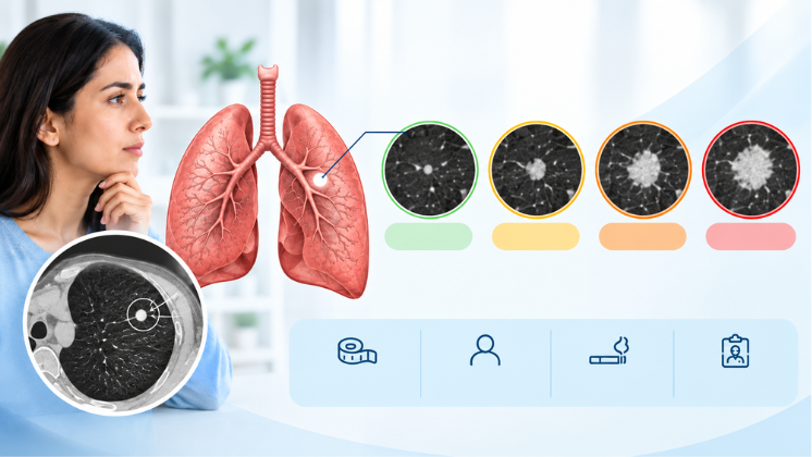
A lung nodule is a small, round spot in the lung that shows up on a chest X-ray or CT scan. Doctors call it a nodule when it is up to 3 cm across. Anything larger is called a mass and is assessed differently.
Nodules are very common. The same JAMA review found them on about 30% of chest CT scans. Most people with a nodule have no symptoms. The nodule is simply an incidental finding.
Are lung nodules always cancer?
No. Most are scars or small marks left behind by an old infection or inflammation. Common causes include:
- Healed infections: old tuberculosis, fungal infections or pneumonia can leave a small, often calcified, scar called a granuloma.
- Active infection or inflammation: some nodules are a current infection and shrink or disappear on the next scan.
- Lymph nodes inside the lung: small and harmless, with a typical shape radiologists recognise.
- Benign growths: such as a hamartoma, which is non-cancerous.
- Lung cancer or a spread from cancer elsewhere: this is the minority, but it is why some nodules need follow-up.
In India, tuberculosis adds a layer most Western guidelines don’t account for. According to the WHO Global Tuberculosis Report 2025, India had 25% of the world’s TB cases in 2024. Many adults here carry small healed TB scars without ever knowing they had TB. The flip side is just as important. A nodule should not be assumed to be TB and treated with TB medicines without proof, because lung cancer can look similar on a scan. Getting a sample when it’s needed is how the two are told apart.
How many lung nodules are normal?
There isn’t a “normal” number, but several small nodules are usually less worrying than a single larger one. Multiple tiny nodules often point to a past infection. For multiple small ground-glass nodules (under 6 mm), the Fleischner Society guidelines specifically suggest considering an infection as the cause. What matters for each nodule is its size, its type and whether it changes over time.
What size lung nodule is concerning?
Size is the single strongest clue. The guidance doctors commonly use for nodules found incidentally on CT comes from the Fleischner Society. It applies to adults aged 35 and above who are not being screened for lung cancer, do not have a known cancer and are not immunocompromised.
The guidelines split people into low risk and high risk. Higher risk means features such as a long smoking history, older age, a family history of lung cancer, emphysema or lung scarring, or a nodule in the upper part of the lung with irregular, spiky edges. Your doctor weighs these together with the nodule’s size.
Solid nodules: size, cancer risk and next step
| Nodule size | Chance of cancer | Low-risk adult | High-risk adult |
|---|---|---|---|
| Under 6 mm | Less than 1% | No routine follow-up | Optional CT at 12 months if shape or location is suspicious |
| 6–8 mm | About 1–2% | CT at 6–12 months; possibly one more at 18–24 months | CT at 6–12 months and again at 18–24 months |
| Larger than 8 mm | Depends on shape, growth and risk factors | Consider CT at 3 months, PET-CT or a biopsy | Consider CT at 3 months, PET-CT or a biopsy |
Cancer risk figures are from a 2022 JAMA review. Follow-up steps are from the 2017 Fleischner Society guidelines, the most recent edition.
For scale: 6 mm is roughly the size of a pencil eraser, and 8 mm about the size of a small pea.
Ground-glass and part-solid nodules
Some nodules look hazy rather than solid on the CT. These are called ground-glass nodules, or part-solid if they have a solid core inside the haze. They need a different approach because some early, slow-growing lung cancers look like this.
- A pure ground-glass nodule of 6 mm or more is rechecked at 6–12 months, then every 2 years up to 5 years.
- A part-solid nodule of 6 mm or more is rechecked at 3–6 months. If its solid part is 6 mm or larger, or it is growing, PET-CT or a biopsy is considered.
- The JAMA review puts the chance of cancer at 10–50% for ground-glass nodules larger than 10 mm that are still there after 3 months.
How fast can a lung nodule grow, and can it go away?
This is why the follow-up CT matters more than any single scan.
- It shrinks or disappears: it was most likely inflammation or infection. This is one reason the guidelines ask for a repeat scan before acting on many nodules.
- It stays exactly the same: this is reassuring. For solid nodules, the Fleischner guidelines end follow-up at around 18–24 months if nothing changes.
- It grows or develops a solid part: this is the signal to move from watching to testing.
Ground-glass nodules can grow very slowly, which is why their follow-up runs to 5 years rather than 2.
One practical point: always compare like with like. Ask for your earlier CT films or the CD, not just the printed report, so the new scan can be compared measurement for measurement.
When does a lung nodule need a biopsy?
A biopsy is considered when the chance of cancer is high enough that waiting would be the bigger risk. In practice that usually means one of these:
- A solid nodule larger than 8 mm with a worrying shape or risk profile.
- A nodule that has grown on a follow-up CT.
- A part-solid nodule whose solid core is 6 mm or more, or is getting bigger.
- A nodule together with enlarged lymph nodes in the centre of the chest.
- A PET-CT that shows the nodule is metabolically active.
A note on PET-CT in India. PET-CT highlights cells that use a lot of glucose. Cancer cells do, but so do cells fighting an active infection. In a country with a high TB burden, a nodule that “lights up” on PET can still turn out to be tuberculosis. A 2020 report in BMC Medical Imaging described TB lesions with PET uptake similar to cancer. So PET-CT guides the decision, but it can’t replace a tissue diagnosis.
How is a lung nodule biopsy done?
The right method depends on where the nodule sits and whether lymph nodes are involved. The aim is the safest route that gives a reliable sample, ideally one that can also stage cancer at the same time if it is found.
| Method | Best suited for | What it involves |
|---|---|---|
| Bronchoscopy with biopsy and BAL | Nodules near the larger airways; suspected infection, including TB | A thin flexible camera through the nose or mouth under sedation; day-care |
| EBUS-TBNA | Enlarged lymph nodes in the centre of the chest; staging lung cancer; TB or sarcoidosis in lymph nodes | Bronchoscope with an ultrasound tip; a fine needle samples nodes through the airway wall, without a surgical cut; day-care |
| Radial EBUS-guided bronchoscopy | Smaller nodules further out in the lung | A miniature ultrasound probe helps locate the nodule from inside the airway before sampling |
| CT-guided needle biopsy | Nodules close to the chest wall | A needle passed through the skin under CT guidance, usually done by an interventional radiologist |
| Surgical removal | Highly suspicious nodules where removing it is both the diagnosis and the treatment | Keyhole (VATS) surgery by a thoracic surgeon |
Bronchoscopic methods have one practical advantage in India. The same sample can be sent for cancer pathology and for TB tests together, so one procedure can answer both questions. EBUS can also stage the lymph nodes in the same sitting if cancer is found.
Dr. Kishan Srikanth Juvva holds a Master’s in Interventional Pulmonology from the University of Florence, Italy, with training in flexible bronchoscopy and EBUS (linear and radial). These procedures are performed at Apollo Hospitals, Financial District, where he works alongside the hospital’s oncology, radiology and thoracic surgery teams when a nodule needs more than one specialist.
When to see a pulmonologist about a nodule
A nodule doesn’t usually need an emergency visit, but it does need a plan. See a pulmonologist if:
- Your CT report mentions a nodule and nobody has told you whether or when to repeat the scan.
- The nodule is 6 mm or larger, or it is hazy (ground-glass) or part-solid.
- A follow-up scan shows any change in size or appearance.
- You smoke or used to smoke, have a family history of lung cancer, or have worked around dust, asbestos or fumes.
- You have a cough that isn’t settling, are coughing up blood, or are losing weight without trying.
- You were started on TB treatment for a nodule without a sample confirming TB.
If you’re unsure what your report means, a second opinion on your chest CT can settle it. A pulmonologist looks at the images themselves, not just the report, and gives you a clear plan with dates.
Getting a lung nodule assessed in Hyderabad
Dr. Kishan Srikanth Juvva is a pulmonologist in Hyderabad and Senior Consultant in Clinical & Interventional Pulmonology at Apollo Hospitals, Financial District. His training:
- MD from Guntur Medical College
- DM in Pulmonary & Critical Care Medicine from VMMC & Safdarjung Hospital, New Delhi, after securing All India Rank 2 in NEET Super Speciality
- Master’s in Interventional Pulmonology from the University of Florence, Italy
- Fellowship in Sleep Medicine from ISDA, New Delhi
| Apollo Hospitals, Financial District | Breathe Well Clinic, Kondapur | |
|---|---|---|
| Best for | Assessment, biopsy (bronchoscopy, EBUS) and multi-specialty planning | Evening consultation and review of CT reports |
| Timings | Monday–Saturday, 9:00 AM–5:00 PM | Monday–Saturday, 5:00–8:30 PM |
| Address | 1st Floor, Room 3, Apollo Hospitals, Financial District, Nanakramguda | 1st Floor, Kadiri’s Apurupa Urban Apartments, Botanical Garden Road, beside Aparna Heights 1, Kondapur |
| Convenient from | Gachibowli, Kokapet, Narsingi, Manikonda, Jubilee Hills | Kothaguda, Madhapur, Hitech City, Hafeezpet, Miyapur |
| Book | +91 90598 73547 | +91 87122 23696 |
Bring with you: the CT films or CD (not only the printed report), every earlier chest scan you have, any PET-CT, and a list of your medicines. If you’ve had TB treatment in the past, bring those records too.
Frequently asked questions
1. Is a 4 mm lung nodule dangerous?
A solid nodule under 6 mm has less than a 1% chance of being cancer. In adults at low risk, the Fleischner guidelines recommend no routine follow-up. If you smoke or have other risk factors, your doctor may suggest one CT at 12 months.
2. Can a lung nodule cause cough or chest pain?
Usually not. Most nodules are too small to cause symptoms and are found by chance. If you have a persistent cough, chest pain or are coughing up blood, that needs assessment in its own right, whatever the nodule’s size.
3. Can lung nodules go away with antibiotics?
Nodules caused by an active infection can shrink or disappear, sometimes after treatment. That is one reason a repeat CT is often advised before any biopsy. Antibiotics should not be taken “just in case” without a doctor’s assessment.
4. Should a lung nodule be treated as TB?
Not without proof. TB is common in India, but lung cancer can look similar on a CT. If TB is suspected, a sample from bronchoscopy or EBUS can confirm it before treatment starts.
5. Does a nodule that lights up on PET-CT mean cancer?
Not necessarily. Infections, including TB, can also show uptake on PET-CT. A biopsy gives the answer.
6. Is bronchoscopy or EBUS painful?
Both are done under sedation with local anaesthetic to the throat, so most people feel little or nothing. Most go home the same day. Your doctor will explain preparation, such as fasting, beforehand.
Medically reviewed by Dr. Kishan Srikanth Juvva, MD, DM (Pulmonary & Critical Care), Senior Consultant, Apollo Hospitals, Financial District, Hyderabad. This article is for general information and does not replace a consultation with your doctor.
Sources
- Mazzone PJ, Lam L. Evaluating the Patient With a Pulmonary Nodule: A Review, JAMA, January 2022
- MacMahon H, et al. Guidelines for Management of Incidental Pulmonary Nodules Detected on CT Images: From the Fleischner Society 2017, Radiology, 2017
- World Health Organization, Global Tuberculosis Report 2025, as reported by Scroll.in, November 2025
- FDG PET/CT findings in tuberculosis mimicking malignancy, BMC Medical Imaging, 2020